
When Is a Biopsy Necessary? Understanding Image-Guided Biopsy
Finding an abnormal lump, swelling, mass, or suspicious area on an ultrasound, CT scan, or MRI does not automatically mean cancer. Many abnormalities are benign, inflammatory, or related to infection. However, imaging alone cannot always determine exactly what an abnormal tissue represents.
In such situations, a doctor may recommend a Biopsy Testing Centre In Jaipur. A biopsy involves collecting a small tissue sample from the suspicious area so that it can be examined under a microscope by a pathologist.
Modern image-guided biopsy allows doctors to target abnormalities accurately using ultrasound, CT, or other imaging techniques, often without the need for open surgery.
What Is anImage-Guided Biopsy?
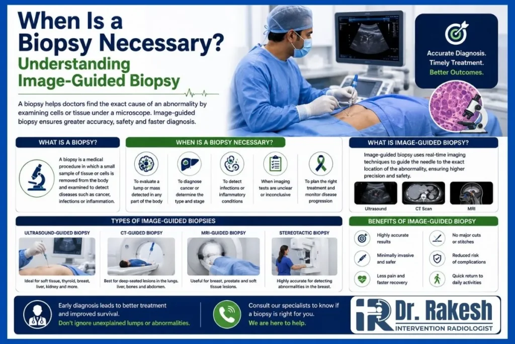
An image-guided biopsy is a minimally invasive diagnostic procedure in which medical imaging is used to guide a needle into a specific abnormal area.
Depending on the location of the lesion, guidance may include:
• Ultrasound
• CT scan
• Mammography for selected breast lesions
• Other specialized imaging techniques
The imaging allows the doctor to visualize the target and guide the biopsy needle toward the appropriate tissue.
This approach is particularly useful when an abnormality is small, deep inside the body, or located close to important organs and blood vessels.
When Is a Biopsy Necessary?
A biopsy may be recommended when clinical examination or imaging identifies an abnormality that cannot be confidently diagnosed through imaging alone.
Common situations include the following.
1. When a Lump Has Suspicious Features
Many lumps are harmless, but some require tissue examination.
A doctor may recommend biopsy when a lump:
• Continues to increase in size
• Feels unusually hard or fixed
• Persists for a long period
• Has suspicious imaging characteristics
• Returns after previous treatment
• Cannot be clearly diagnosed through other investigations
A biopsy provides actual tissue that can be examined microscopically.
2. When Imaging Shows an Abnormal Mass
Ultrasound, CT, or MRI can identify abnormal areas in organs and tissues, but imaging cannot always establish the exact diagnosis.
For example, a scan may detect a lesion in the liver, lung, kidney, lymph node, breast, or another location.
When imaging findings require confirmation, an image-guided biopsy can obtain tissue directly from the suspicious area.
3. When Cancer Needs to Be Confirmed or Ruled Out
Biopsy Testing Centre In Jaipur is commonly used when doctors need to determine whether abnormal tissue contains cancer cells.
However, being advised to undergo a biopsy does not mean you definitely have cancer.
The purpose is to obtain enough information to distinguish between possibilities such as:
• Benign growths
• Inflammatory conditions
• Infections
• Precancerous changes
• Malignant tumors
The pathology result can then help guide further treatment decisions.
4. When FNAC Does Not Provide Enough Information
FNAC (Fine Needle Aspiration Cytology) collects individual cells or small groups of cells using a fine needle.
It can provide valuable diagnostic information for many lumps and nodules. However, FNAC does not always provide enough information about how cells are arranged within the tissue.
A core needle biopsy collects a small cylinder of tissue, preserving more of the tissue architecture.
Therefore, a biopsy may be recommended when FNAC results are:
• Inconclusive
• Suspicious
• Inadequate
• Unable to provide the information required for diagnosis
FNAC and biopsy are complementary diagnostic techniques rather than competing tests.
5. When Treatment Depends on the Exact Diagnosis
Different diseases can look similar on imaging but require completely different treatments.
For example, treatment for an infection is very different from treatment for an inflammatory condition or malignant tumor.
A tissue diagnosis can help doctors understand the nature of the abnormality before deciding on treatment.
In cancer care, biopsy samples may sometimes provide additional information about tumor type and biological characteristics that can influence treatment planning.
How Does Image Guidance Improve a Biopsy?
Without imaging, a superficial lump that can be easily felt may sometimes be sampled directly.
However, many abnormalities cannot be accurately located by touch alone.
Image guidance allows the specialist to:
• Identify the exact target
• Monitor needle position
• Select an appropriate biopsy pathway
• Avoid important nearby structures
• Sample a specific part of the lesion
• Improve the likelihood of obtaining useful tissue
This makes image guidance particularly valuable for deeper or smaller lesions.
What Happens During an Image-Guided Biopsy?
Before the procedure, your doctor reviews previous imaging, medical history, medications, and relevant blood tests.
The skin is cleaned, and local anesthesia is commonly used to numb the area.
Using ultrasound or another imaging technique, the specialist identifies the target and carefully guides a biopsy needle into the abnormal tissue.
One or more small samples may be collected.
The samples are then sent to a pathology laboratory for microscopic examination.
How Should You Prepare for a Biopsy?
Preparation depends on the type and location of the Biopsy Testing Centre In Jaipur.
Before your appointment, inform your doctor about:
• Blood-thinning medications
• Bleeding disorders
• Allergies
• Existing medical conditions
• Current medicines
• Previous biopsy results
Do not stop blood-thinning medication unless specifically instructed by your treating doctor.
You should also carry previous ultrasound, CT, MRI, FNAC, and other relevant reports.
Is an Image-Guided Biopsy Painful?
Local anesthesia is commonly used, so many patients experience pressure or brief discomfort rather than significant pain.
Some soreness, tenderness, or bruising may occur afterward.
Recovery varies depending on the location and type of biopsy. Your healthcare team will provide specific aftercare instructions.
Are There Risks?
Image-guided biopsy is generally minimally invasive, but no medical procedure is completely risk-free.
Possible risks can include bleeding, infection, discomfort, or injury to nearby structures. Certain biopsy locations have additional procedure-specific risks.
Image guidance and careful patient selection help specialists plan a safe route to the target.
When Will Biopsy Results Be Available?
Biopsy results are not usually immediate because the tissue must be processed and examined by a pathologist.
Some samples may also require special stains, immunohistochemistry, molecular testing, or other additional studies.
Your doctor should interpret the final pathology report together with imaging and clinical findings.
Why Accurate Tissue Diagnosis Matters
The purpose of biopsy is not simply to detect cancer. It is to help establish an accurate diagnosis.
Treating a condition before understanding its nature may lead to unnecessary or inappropriate therapy. A properly performed biopsy can provide information that helps doctors choose the next appropriate step.
Frequently Asked Questions (FAQs)
1. Does being advised to have a biopsy mean I have cancer?
No. Biopsies are performed to determine the nature of abnormal tissue, which may be benign, inflammatory, infectious, or malignant.
2. What is the difference between FNAC and biopsy?
FNAC primarily collects cells, while a core biopsy collects a small piece of tissue, allowing the pathologist to examine tissue architecture.
3. Why is ultrasound or CT used during a biopsy?
Image guidance helps the specialist locate the abnormality and accurately guide the needle toward the intended target.
4. Is an image-guided biopsy a major surgery?
Usually not. Most needle biopsies are minimally invasive and are performed through a small needle puncture rather than a large surgical incision.
5. When might a biopsy be needed after FNAC?
A biopsy may be recommended when FNAC Testing Centre In Jaipur is inconclusive, inadequate, suspicious, or when additional tissue information is required for diagnosis.